Medical technology has been improving at an incredible rate for the past several decades, leading to leaps and bounds in the quality of care for people around the United States. This is especially true for the dental industry.
One of the most important and exciting developments in dental diagnostic technology in the past decade has been the advent of 3D dental imaging. However, most people likely aren’t even aware of what 3D dental technology is — that’s how new it is!
What is 3D dental imaging? Why is 3D dentistry better, and how does this affect you, the patient?
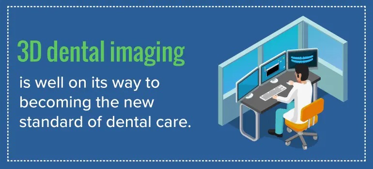
The short answer is this: 3D dental imaging is well on its way to becoming the new standard of dental care. This incredible technological advancement allows dentists to see your mouth in a way never previously possible with X-ray or medical CT scan technology. 3D dental imaging allows them to see your teeth and skull, diagnose any problems and provide more effective treatment for you.
To better understand what 3D dental imaging is, what it has done for the dental industry and what it can do for you and your dental care, we’ll walk you through the incredible machine that makes it possible.
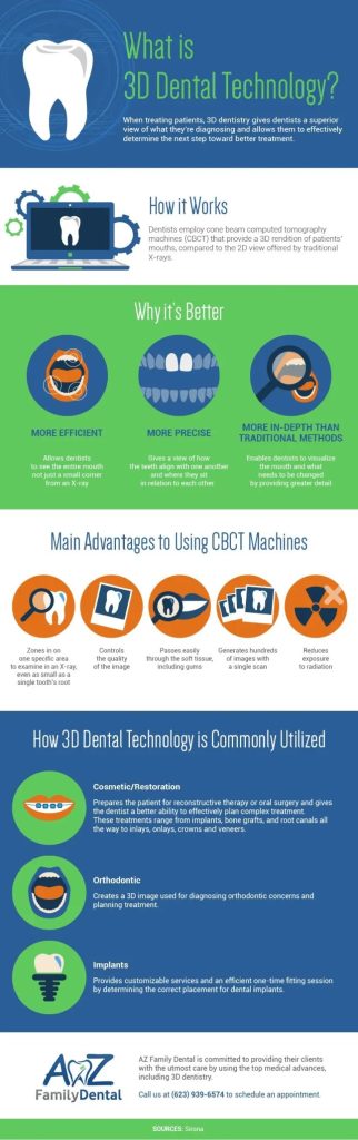
What Is 3D Dentistry?
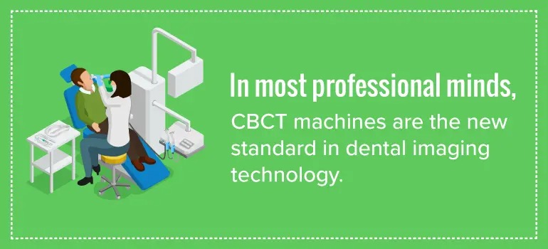
When we think about visiting the dentist’s office, we often think of bright lights, blue bibs, toothbrushes, picks, drills and bubblegum toothpaste. However, advanced imaging utilizing CBCT technology is slowly but surely making its way into dental offices across the United States. Because of their wide recognition as contributors to quality dental care, CBCT machines are now becoming more commonplace among technologically up-to-date offices across the world.
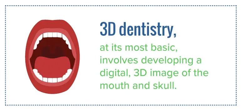
3D dentistry, at its most basic, involves developing a digital, 3D image of the mouth and skull. Traditional X-rays only develop a 2D picture of your mouth, while other methods took longer bouts of radiation to create a detailed picture, and were more unpleasant and intrusive to the patient.
3D dental images are most often used for diagnosis and treatment planning. Being able to see the mouth in three dimensions allows the dentist to better and more effectively formulate an approach to treat dental conditions.
What Are the Advantages of 3D Dentistry?
The unique way 3D dentistry works means it offers numerous features found nowhere else in dental technology. This technology has a huge number of advantages over technologies like traditional X-ray and medical CT scans. The following are some of the benefits of 3D dentistry from a diagnostic standpoint:
- Beam Limitation: The size of the primary X-ray beam in 3D CBCT scanners limits the radiation to only the area of interest, reducing the patient’s exposure to radiation as much as possible.
- Short Scan Time: Because the CBCT scans involved in 3D dentistry can acquire all scan images in a single rotation, scan time isn’t much different from a normal scan. This reduces the chance of image defects due to the natural movement of the patient. It also minimizes time spent on the scan.
- Image Accuracy: 3D CBCT imaging has an incredible resolution, allowing for highly accurate imaging and measuring. This means these images can be used to pinpoint the exact location for a dental procedure.
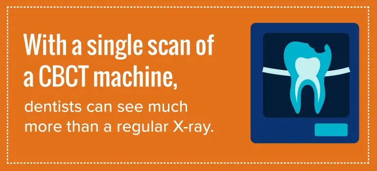
- Image Detail: With a single scan of a CBCT machine, dentists can see much more than a regular X-ray. They can see pathology, infections, nerves, musculature and so much more. This helps dentists see and properly treat dental-caused sinus issues and plan for root canals, implants and extractions. The possibilities are endless with 3D CBCT scans.
- Bone Quality Assessment: 3D CBCT scans can also be used to assess bone quality, which is an important part of evaluating the presence of sufficient bone for implant placement. It is also helpful in determining the size and location of lesions and breaks.
- User-Friendly: 3D dental equipment is very easy to learn, and it’s designed for a trained dentist or dental technician to learn and work with easily.
- Interactive Display: The most important benefit for dental practitioners is the unique ability to demonstrate features in 3D that traditional imaging cannot. The practitioner can reorganize data and magnify and annotate the image.
But what are the advantages of using 3D dentistry for the patient? As it turns out, the benefits of this machinery are just as extensive for patients as they are for dentists. Some of the benefits of 3D dentistry for patients include the following:
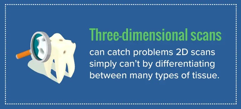
- Diagnostic Accuracy: Three-dimensional scans can catch problems 2D scans simply can’t by differentiating between many types of tissue. Pathology, infections, and abnormal sinus anatomy and joint dysfunctions can all be properly visualized and identified with 3D CBCT imaging. This means patients are properly diagnosed the first time and can get appropriate help much sooner than they would with previous methods.
- Minimal Radiation Exposure: Repeated prolonged exposure to radiation can cause eye damage, the development of malignancies and other health risks, which is why new medical technologies seek to reduce patient exposure. When compared to traditional medical CT scans, 3D CBCT scans emit substantially less radiation, reducing the dosage up to 98.5%.
- Non-Intrusive: No need to bite down on a mold or piece of plastic. The CBCT can scan your entire head without you needing to do anything. This is especially helpful for patients with particularly sensitive gums or teeth, as well as pediatric patients.
- Lower Cost: Going to a third-party imaging center for a medical CT scan can be extremely expensive. Bringing in a 3D CBCT dental imaging device provides all the technology into one place, eliminating the middle-man and saving you money.
- Short Scan Time: A typical 3D CBCT scan takes around ten seconds to complete, meaning your dentist can see and solve problems more quickly than ever. An in-house 3D CBCT scanner at your dentists’ office also means you don’t need to go to a third-party imaging company for a medical CT scan.
A third-party imaging company will likely make you wait days before scheduling your scan, and it could take another few days for them to send your results to your dentist, unnecessarily prolonging the process. All of this can be avoided with an in-house dental 3D CBCT machine.
How Does 3D Dentistry Work?
The 3D dental imaging process starts with taking a scan of the lower half of the face to create an image. The most widely recognized and technologically advanced method of completing this scan is with a cone-beam computed tomography machine, also known as a CBCT machine. This machine takes a scan of the mouth using a series of small beams of radiation, each of which produces a digital image. This series of images is formed, collected and compiled, at which point they can be converted into a three-dimensional model and used for a variety of dental purposes and procedures.
3D dentistry can be used for simple diagnostics, showing angles and features of the teeth that may not have been visible with a traditional 2D X-ray scan. However, this 3D CBCT scan can also be used to develop a complete 3D model of the skull and teeth, which can be used for complex diagnostics and comparative data. This is especially important for identifying degenerative conditions or potential problem areas down the road.
In addition to this diagnostic use, 3D dental imaging can now be integrated with digital impressions of teeth. CEREC technology allows a dentist to take digital impressions of the teeth with extreme accuracy. These digital impressions, which are used to create dental restorations, can then be merged with the 3D CBCT scan. This allows for a seamless planning of complex treatment such as dental implants, customized sleep appliances and corrective devices. This level of integration utilizing CEREC and CBCT is unmatched in the dental industry.
What Is a CBCT Machine?
The cone-beam computed tomography machine, or the CBCT machine, is widely regarded as the new pinnacle of 3D imaging technology, and it represents the official shift from 2D imaging to 3D imaging. This machine uses a cone-shaped X-ray beam to take pictures of the skull and teeth, and then it uses those pictures, along with robust digital processing software, to reconstruct a 3D image. Provided the settings are correct and the patient does not move during the image-collecting process, this image is extremely accurate, with few, if any, distortions.
Today, cone-beam computed tomography is used in the fields of dentistry, radiotherapy and mammography, among others, and it’s used as a safer and more effective alternative to medical CT scans. The number of dental practices, dental and otherwise, using CBCT units in North America and Europe is experiencing a steady rise as more people discover the incredible benefits of CBCT technology. This growth is only hindered by the cost of implementing the system.
Along with this steady growth of implementation, professionals around the world are in the process of developing new standards and applications for this technology. Currently, worldwide dentistry organizations are developing standards of practice for using CBCT units safely, while others are theorizing potential new uses for the technology in science and medical research.
This is primarily due to the unique way in which these machines work.
How Does a CBCT Machine Work?
The CBCT machine is essentially a digital X-ray scanner mounted on a rotating arm. The machine uses a rotating X-ray source, which emits a cone-shaped beam of radiation through the head, or alternative area of interest, onto an X-ray detector on the opposite side. The X-ray source and detector rotate around a fulcrum located directly above the patient, positioned so the head is at the center of the system. The system takes anywhere from 150 to 600 projection images over the course of a few seconds, taking the images in either a complete or partial arc, depending on the focus.
A more detailed description of the process is as follows:
- Situating the Patient: Depending on the type of cone beam imaging system used, the subject may be positioned in a supine, standing or sitting position, with the head or area of interest placed at the center of the CBCT system. Of these positions, sitting is most common, as it is most comfortable and sustainable for the patient and is more accommodating for disabled patients.Upon situating the patient in place, the patient’s head will be stabilized with a restraint mechanism, such as a chin rest, to minimize movement during the scanning process.
- Acquiring the Scan: The X-ray source rotates around the head, while a reciprocating area detector moves opposite, both rotating around a fixed fulcrum oriented directly over the patient’s head. The frame rate, speed of rotation, field of view and completeness of the trajectory arc are all set to get the image desired by the dental practitioner. During this process, the machine emits radiation in short bursts, meaning the exposure time of the patient is only a fraction of the total scan time. This scan takes about ten seconds on average.
- Detecting the Image: The detector picks up the radiation emitted from the X-ray and collects it as digital data. This data is then transmitted to and collected at a dedicated computer.
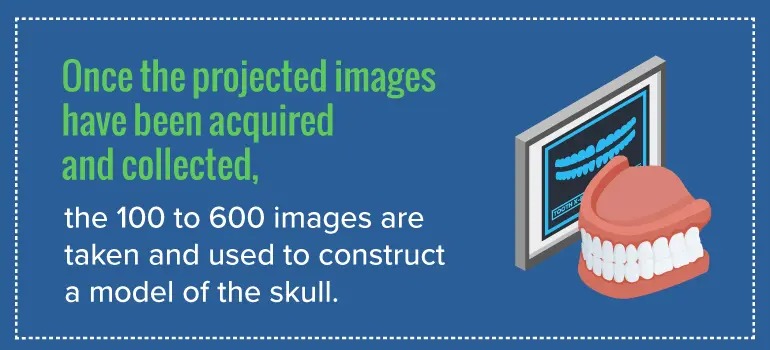
- Reconstructing the Image: Once the projected images have been acquired and collected, the 100 to 600 images are taken and used to construct a model of the skull. This is done with a software that uses reconstruction algorithms to build up a three dimensional model of the skull, teeth or other area of interest. This can take several minutes to complete, depending on the quality of the software and computer hardware.
- Displaying the Image: CBCT technology provides a complete digital model at the end of the process. The software also provides the dental clinician with a relatively large choice of display formats, allowing for 2D, 3D and panoramic views of the mouth and head, along with other viewing options to help focus in on areas of interest.
This unique process results in an extremely useful digital model, which can be referenced for diagnostic and treatment purposes. It also sets it apart from previous methods as a more advanced option.
How Is CBCT Different From Other Methods?
The CBCT process is quite different from a medical CT scan or a traditional X-ray, primarily due to the way in which the images are collected and processed.
Medical CT scans use a fan-shaped X-ray beam that runs through the body, and they take these scans in a helical progression throughout the area of interest to develop individual slices of the image in the field of view. This may require multiple passes to get the same image a CBCT machine could get in one, making the CT scan more expensive to use and increasing the patient’s exposure to radiation.
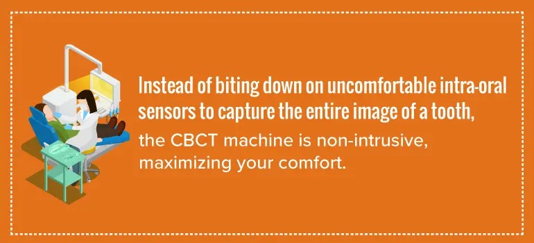
CBCT scans also differ from the traditional dental X-ray. Instead of biting down on uncomfortable intra-oral sensors to capture the entire image of a tooth, the CBCT machine is non-intrusive, maximizing your comfort. Additionally, typical dental X-rays only focus on your teeth, and not on your entire skull or jaw. For more extensive dental surgery or implants, this is simply insufficient. Also, each traditional X-ray picture requires exposure, increasing the time between taking the X-ray and gleaning any meaningful information from it.
Why Should You Get a Dental CBCT Scan?
The benefits of using a CBCT machine are all indicative of a welcome advancement to dental technology. However, the real question you may ask yourself is how 3D dentistry can improve your dental health.
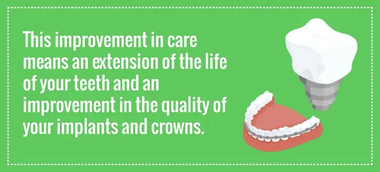
3D dentistry can provide you with top-of-the-line care previously unheard of for the general population, with imaging that makes dental examination, diagnosis and care much faster and effective, both for you and your dentist. This improvement in care means an extension of the life of your teeth and an improvement in the quality of your implants and crowns.
Why Choose AZ Family Dental?
When it comes to dental technology, AZ Family Dental prides itself on being at the forefront. Our fully functional 3D imaging systems are top-of-the line and are ready to help you with your dental needs.
We are proud to be at the forefront of 3D dentistry with the Orthophos SL-AI. This complete X-ray solution is a non-intrusive CBCT X-ray at the height of 3D imaging technology. Its sharp image quality and incredible imaging software allow 2D and 3D images to be taken with ease.
Outside of our technological focus, AZ Family Dental has provided the Glendale, Peoria and Greater Phoenix areas with the best family, cosmetic and dental implant services around. We have a long and steady history of quality care at a fair price, and we strive every day to maintain that reputation. From your child’s first cleaning to your next root canal, we will be there for you and your family, offering the best service and expert care.
Is your family looking for a quality dentistry service on the cutting-edge of technology? Do you want to learn more about 3D dental imaging? Contact AZ Family Dental today to learn what services we can offer you and your family.